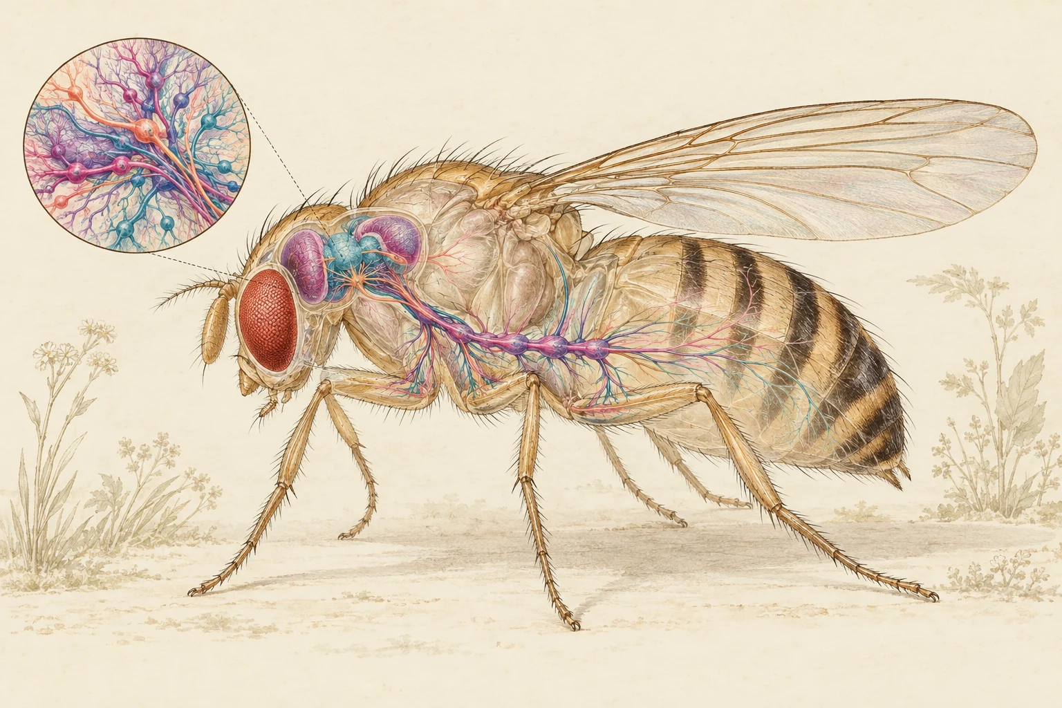
A fruit fly’s nervous system offers a way to study how sensation becomes action, one connection at a time. On September 3, 2026, an HHMI Janelia-led collaboration reported a male fly connectome containing more than 166,000 neurons across the brain and ventral nerve cord. Google Research’s account of the collaboration describes how the map links sensory pathways with the circuits controlling the body.
What the map records
A connectome is a wiring diagram: it records neurons and their connections. Electron microscopy, computational reconstruction and human proofreading turn tissue images into a navigable network. The earlier 2024 FlyWire female brain map contained 139,255 neurons and 54.5 million chemical synapses between them. Its companion cell atlas helps researchers identify and compare cell types. Those counts describe a different specimen and scope, not a smaller version of the same map.
From wiring to experiments
Connectivity becomes especially useful when it produces a prediction. In a 2024 study by Shiu and colleagues, a simplified computational model used FlyWire connections and predicted neurotransmitters to identify neurons involved in feeding and grooming. The researchers checked selected predictions in living flies, including by activating neurons and observing behavior.
Why it is not a complete digital brain
A map alone does not reproduce learning, hormones or every detail of neural activity. Eon’s virtual-fly technical report describes simplified neurons, limited behaviors and approximate connections between brain and body models. The practical advance is a better starting point for experiments: researchers can propose a circuit explanation, test it, and revise the model when the fly behaves differently.
Sources reviewed September 24, 2026. This article summarizes published maps and models; it does not independently validate them.